Increase throughput as needed.
Achieve unattended operation, night operation or full process automation, significantly reducing manual operations and improving efficiency.
More consistent orifice handling for more reliable results.
Better environmental control maintains good cell status.
preface:
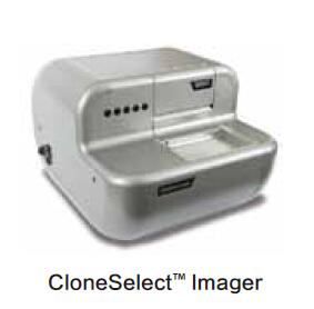
Obtaining a stable, high-yielding cell line often requires screening thousands of cell clones. In addition to finding these rare, high-quality cell lines, researchers need to prove that they are derived from individual cells (ie, monoclonal) to meet FDA requirements. Therefore, researchers need to spend a lot of time and effort to screen single-cell clones. To alleviate this burden, Molecular Devices has developed the CloneSelect Imager Cell Imaging Analysis System, which automatically captures images of individual cells and monitors their growth. Here we show three examples of integrating CloneSelectImager with automated processes to increase detection throughput.
Example 1 is a simple automation solution that meets the throughput requirements of thousands of clones per day. Example 2 allows for longer monitoring (typically 2-3 weeks) for automatic monitoring. Example 3 integrates clonal growth monitoring and product analysis.
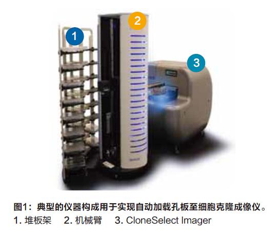
Experiment 1: Automatic loading of the plate to the cell clone imager
Instrument composition: CloneSelect Imager, robotic arm and stacker
Although there are a variety of methods for demonstrating monoclonality, the method of providing single-cell images is relatively simple and clear, and is an important method. However, collecting single-cell images of thousands of clones is an extremely labor-intensive process, especially in the case of microscopic observation. CloneSelect Imager can be used to quickly image the entire well plate, determine if there is only one cell in each well, and monitor its subsequent growth. CloneSelect Imager can only image one plate at a time when used independently, which requires manual insertion into the plate. However, by integrating the CloneSelect Imager with a third-party robotic arm, 48 orifice plates can be processed at room temperature (Figure 1), significantly increasing the unguarded run time to over 2 hours.
Example 2: Automatically take the plate from the cell culture box and put it back
Instrument composition: CloneSelect Imager, robotic arm and incubator
Although Case One increases the daily unguarded run time, it is necessary to put the orifice plate into the stack every day and retrieve it at the end. In addition, placing the well plate at room temperature for extended periods of time may affect cell viability. In this case, we connected the CloneSelect Imager to the cell culture incubator via a robotic arm (Figure 2), thereby reducing the effects on cell viability. This not only reduces temperature fluctuations, but also achieves a longer time (2-3 weeks) of unguarded operation, and the user does not need to operate the orifice plate, thereby reducing the risk and difference of contamination. In this example, we integrated a 42-well plate incubator, and we also have a higher capacity incubator that can hold approximately 200 plates.

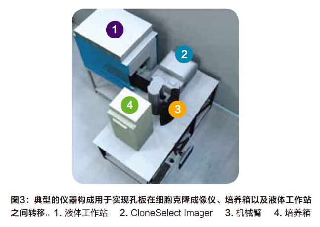
Example III: Analysis of downstream automatically taken from the culture plates forming the box, and the supernatant was drawn
Instrument composition: CloneSelect Imager, robotic arm, incubator and liquid workstation
After monitoring the single cell formation clone, the yield was measured. A variety of assays are available to achieve this, including ELISA for antibodies such as SpectraMax® i3x from Molecular Devices, or label-free methods such as ForteBio's Octet® system. Generally, after the clones are grown to a certain size, the supernatant is taken up for analysis, but considering that the growth rates of the clones are inconsistent, it is a time-consuming process to manually judge and operate one by one. In this automated solution, single cells were seeded into multiwell plates and placed in an incubator, and cell growth was monitored by CloneSelect Imager for the next 2 weeks (Figure 3). During this time, wells with more than 80% cell confluence will be selected for downstream analysis, thus solving the problem of inconsistent clonal growth rates. Beckman Coulter's liquid workstation can aspirate the supernatant of the selected wells and transfer them to a new well plate for downstream analysis.
summary:
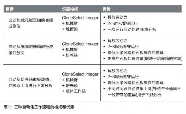
The need to shorten the cell line development cycle is increasing, and the three automation solutions described above can achieve unattended operation, increase detection throughput, and provide the proof materials needed for cell line development (Table 1). In addition to increasing throughput, these automated solutions free users from repetitive work and provide consistent and reliable data.
Can be used directly to maintain the integrity of cell morphology Guidelines recommend cell preservation fluids for sample preservation and transport. It can be used for sputum, bronchoscopic alveolar lavage fluid, pleural fluid and other liquid samples.
Sample storage tubes,Medical disposable storage tubes,Disposable medical consumables
Jiangsu iiLO Biotechnology Co., Ltd. , https://www.iilogene.com