The difference between small animal optical living imaging and small animal photoacoustic imaging system
Author: Di Junhui
Small moving optical in vivo imaging mainly uses bioluminescence and fluorescence. Bioluminescence is the labeling of cells or DNA with the luciferase gene, while fluorescent techniques are labeled with fluorescent reporter groups (GFP, RFP, Cyt and dyes, etc.). Using a very sensitive optical inspection instrument, researchers can directly monitor cellular activity and genetic behavior in living organisms. Due to its extremely simple operation, intuitive results, and high sensitivity, it has been widely used in life sciences, medical research, and drug development in the few years since its development.
Small animal photoacoustic in vivo imaging is a non-destructive medical imaging method developed in recent years. It combines the high contrast characteristics of optical imaging with the high penetration depth of ultrasound imaging to provide high resolution and high contrast tissue imaging. The purchase of this system fully considers the needs of scientific research and practical application, and can carry out cardiovascular diseases (angiogenesis, myocarditis, thrombosis, myocardial infarction, etc.), lymph, tumor, nervous system, blood disease, new molecular exploration for small animals. The cutting-edge research on needle, hemoglobin concentration and blood oxygen saturation measurement and functional imaging will further enhance the research level and status of scientific research units in these fields. Photoacoustic technology has better bio-tissue penetration than near-infrared technology, and also has high resolution and no side effects, and is gradually becoming another research hotspot in the field of non-destructive testing of biological tissues.
Since both the optical living body and the photoacoustic living body can perform living body imaging in small animals, there may be confusion about these two types of techniques. Let us look at the difference between the two.
1. Imaging mode.
Optical live imaging mostly uses a 2-D imaging mode, resulting in a planar two-dimensional image. The image does not have depth and depth information cannot be obtained, so the animal organs are stacked together and the source of the fluorescent signal cannot be determined exactly. The photoacoustic imaging adopts the 3-D imaging mode, and the stereoscopic three-dimensional structure image is obtained, and the angle information can be arbitrarily cut and viewed from the three-dimensional direction. The true 3-D imaging mode has become the development trend of molecular imaging products, so from this point of view, photoacoustic imaging is in line with the development of molecular imaging.
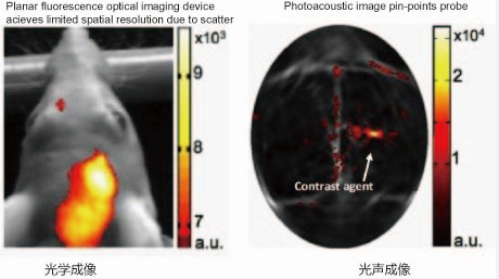
2. Resolution.
Optical in vivo imaging typically has micron-scale optical planar resolution with no spatial resolution. Photoacoustic imaging has a three-dimensional spatial resolution on the order of micrometers and is isotropic resolution. Isotropic resolution produces higher quality, uniform, and sharp images.
3. Light source.
Optical live imaging typically uses a xenon lamp with a filter to produce different wavelength combinations, and the choice of filter is always accompanied by an increase in cost. Moreover, due to the size of the space, the number of filters cannot be infinitely expanded, and the cost of xenon lamps is lower than that of lasers, but the stability and monochromaticity are poor. Photoacoustic imaging generally uses OPO near-infrared adjustable pulse laser. The laser can be adjusted in 1 nm steps, and can be continuously adjusted from 680-950 nm. The laser energy is stable and the service life is long. Laser-based optical devices are already the first choice for molecular imaging product development.
4. Sensitivity.
Both optical and photoacoustic imaging have a sensitivity of at least nanomolar. However, optical live imaging is accompanied by the reflected signal of the animal's body hair, which is often disturbed, which makes the true sensitivity greatly compromised. The photoacoustic signal benefits from the anti-interference of the near-infrared material, and the sensitivity depends on the ability to distinguish the target signal from the background, so it is superior to the optical imaging device.
5. Anti-interference ability.
Due to the influence of mouse body hairs, optical imaging is often accompanied by very large reflection and scattering signals, and it is impossible to determine the specific parts. Photoacoustic imaging Because the photoacoustic is emitted by the laser, it is detected by ultrasonic, completely avoiding the phenomenon of optical scattering and reflecting light, and can present a very accurate and clear localized positioning image. Photoacoustic is the combination of optical and acoustic advantages, with a penetration depth of up to 7cm, avoiding signal interference, so optical sound has an absolute advantage in anti-jamming.
6. Signal acquisition method .
Optical imaging is acquired using a CCD system to obtain a planar two-dimensional picture. Most of the photoacoustic signals are detected by 128 detectors, and the stereoscopic three-dimensional signal information is obtained.
7. Kinetic studies.
Optical imaging can only obtain planar two-dimensional image information, and photoacoustic can study the three-dimensional distribution of various organs on the target metabolite. One of the advantages of 3D imaging is the ability to perform analyses such as metabolism, pharmacokinetics, and biodistribution.
Therefore, combining the above aspects, we can see that photoacoustic imaging has become a trend in the development of molecular imaging. A series of recent studies have shown that photoacoustic imaging will soon be approaching clinical applications.
For example, the Endra Nexus 128 Photoacoustic Imaging System, the only complete 3-D photoacoustic imaging system on the market, accurately determines the probe's distribution in the tissue, while other photoacoustic systems are based on sliced Scanning system. The complete 3-D photoacoustic imaging system determines the Nexus128's superior spatial resolution, sensitivity, animal processing speed, scanning speed and throughput.
(Endra Company Profile: Endra Corporation of the United States was founded by Enlight Biosciences, a group of seven pharmaceutical companies including Pfizer, Merck, Johnson & Johnson, Abbott, Lilly, Novartis, and Astra. Endra's history of developing optical sounds can be Back in 2001, it has been nine years old, and Endra has been conducting applied research on tumor biology and probe development for more than three years. Endra is committed to a true 3-D imaging system; To date, Endra The Nexus 128 is the most installed and used 3-D imaging system.)
Irrigation Filter,Irrigation Filter System,Irrigation Filter Bunnings,Plastic Pe Material Filter
Shandong Yibiyuan Water-saving Equipment Technology Co., Ltd. , https://www.cnybyjs.com