Release date: 2017-10-25
Every year, 40,000 women die of breast cancer in the United States alone. When cancer is discovered at an early stage, they can often be cured. Mammography is the best alternative diagnostic method, but it also has shortcomings. It often results in false positives, which leads to unnecessary surgery.
Common causes of false positives are called "high-risk" lesions, which appear suspicious on X-rays, and abnormal cells can be seen in biopsies. In this case, the patient usually removes the lesion by surgery; however, the lesion is benign in 90% of the cases. This means that the pain, expensive treatment, and postoperative scars that thousands of women experience each year are unnecessary.
However, the necessary surgery can be well eliminated while retaining the important role of X-ray examination in cancer surveillance. Researchers from the MIT Computer Science and Artificial Intelligence Laboratory (CSAIL), Massachusetts General Hospital (MGH), and Harvard Medical School believe the answer lies in artificial intelligence (AI). They jointly published an article in the recent issue of Radiology. 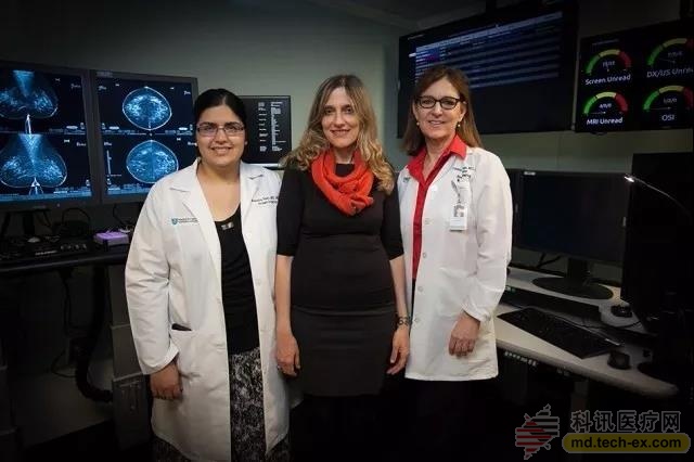
Left to right: Manisha Bahl, Director of the Breast Imaging Scholarship Program at the Massachusetts Hospital; Regina Barzilay, Professor at the Massachusetts Institute of Technology; Constance Lehman, Professor at Harvard Medical School, and Director of the Breast Imaging Department at the MGH Radiology Department. Image source: CSAIL/MIT
The team jointly developed an AI system that uses machine algorithms to predict post-operative mammography and acupuncture biopsy to determine whether high-risk lesions escalate to cancer during surgery. Through the training of more than 600 existing high-risk lesion information, the model looks for patterns in many different data elements, including demographics, family history, past biopsy and pathology reports. Testing 335 high-risk lesions, the model correctly diagnosed 97% of breast cancers as malignant, and can reduce the number of benign surgical procedures by more than 30% compared to current long-term testing methods.
“Because diagnostic tools are so inaccurate, it is an understandable trend for doctors to over-screen breast cancer,†said Regina Barzilay, professor of electrical engineering and computer science at the Massachusetts Institute of Technology, who is also a breast cancer survivor. “When there is a lot of uncertainty in the data, machine learning is the tool we need to improve discovery and prevent over-treatment.â€
"To the best of our knowledge, this is the first study to use machine learning to distinguish between high-risk lesions that require surgery and which are not needed," said Constance Lehman, a professor at Harvard Medical School and director of the MGH Radiology Breast Imaging Division. We believe that we can help women make more informed decisions about their treatment and that we can provide more targeted health care."
So how does machine learning achieve the above goals?
When a X-ray imaging reveals a suspicious lesion, a needle biopsy is used to confirm whether it is cancer. About 70% of lesions are benign, 20% are malignant, and 10% are high-risk lesions.
The way doctors treat high-risk lesions is different. Some have surgery in all cases, while others only perform surgery with a high degree of canceration, such as "atypical ductal hyperplasia" (ADH) or "lobular carcinoma in situ" (LCIS).
The first approach requires patients to experience painful, time-consuming, and expensive surgery, and may be unnecessary; the second is less precise and may result in the omission of some high-risk lesions of cancer outside of ADH and LCIS.
“The vast majority of patients with high-risk lesions do not have cancer, so we try to find those that can be confirmed,†said MGH radiologist Bahl. “In this case, when you try to increase the number of cancers you can recognize. You will also increase the number of false positives you find."
The team used a method called the “random forest classifier.†The team developed a model that avoided unnecessary surgery and always diagnosed more cancer lesions than the strategy of always performing surgery. It is not a strategy to perform surgery only on traditional “high-risk lesionsâ€. In particular, the new model diagnosed 97% of cancers, compared with 79% of traditional methods.
Lehman said: "In the past, we may recommend resection of all high-risk lesions. But now, if the model determines that the lesion has a low probability of cancer in a particular patient, we can discuss the patient's choice in more detail with the patient. For some patients, it is more reasonable that their lesions are based on image removal rather than surgery."
MGH will incorporate this model into clinical practice next year, and the team is working hard to make the model more complete. In the future, this kind of machine learning is expected to be used in more cancer treatment evaluations, which will have more help and reference significance for changing many “one size fits all†practices in traditional models.
Source: Health New Vision
Disposable Mask,Black Disposable Mask,N95 Disposable Mask,Best Disposable Mask
Zhejiang Lanhine Medical Products Ltd. , https://www.lanheyiliao.com